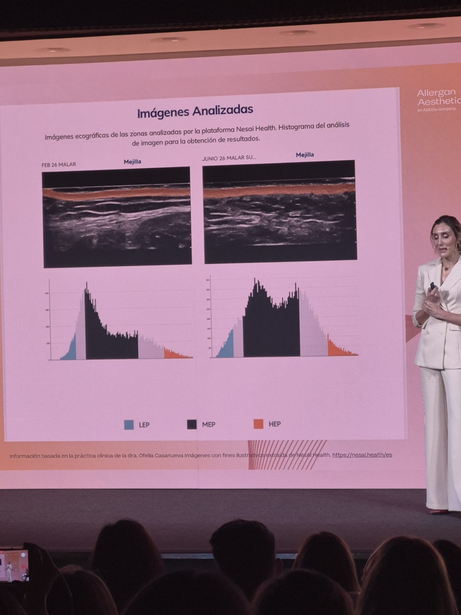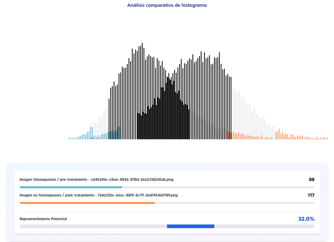
What is the difference between a camera-based skin analyzer and an ultrasound-based one? The difference isn’t the brand or the image sharpness: it is depth. A camera-based analyzer (some are built into cosmetics apps, others into in-clinic devices) photographs the skin surface under different wavelengths and calculates spots, pores, texture or wrinkles. A high-resolution facial ultrasound doesn’t take photographs: it emits ultrasound, which goes through the skin and bounces back differently depending on the density of the tissue it meets. That is why it can “see” what lies beneath the surface, including fillers the patient sometimes doesn’t remember or doesn’t know she has.
They are two different categories of instrument answering different questions. Mixing them up leads you to expect from a surface analyzer something it structurally can’t give you.
The camera sees what light reflects
Camera-based skin analyzers are the most widespread technology in the sector because they are fast, non-invasive and very visual: ideal for a sales consultation or an initial cosmetic assessment. Some examples with different approaches:
| Analyzer | What it sees |
|---|---|
| Vichy Skin Consult AI / Perfect Corp | Surface texture, spots and pores. Geared towards recommending the brand’s own products |
| Antera 3D (Miravex) | 3D surface image: texture, wrinkles, volume. Doesn’t see fillers or inner layers |
| VISIA / OBSERV 520x | Reference multispectral imaging in clinic, without a validated AI report |
| FotoFinder | High resolution for oncological dermatology: a different use case, not aesthetics |
None of them goes through the skin. They all analyse the light reflected back to the camera, not what happens in the deeper layers.
Ultrasound sees the inner structure
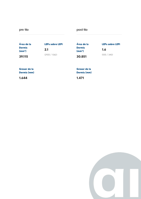
A facial ultrasound with a high-frequency linear probe produces an image of the skin layers (epidermis, dermis, subcutaneous tissue) and of any non-biological material present in them: hyaluronic acid, silicone, calcium hydroxylapatite or biopolymers. On that same image, an AI system can identify and quantify structural parameters that no surface analyzer measures:
- Previous fillers (presence, approximate type, location)
- Dermal thickness and density
- SLEB (subepidermal low-echogenic band)
- Hydration
- Inflammatory processes
- Elastosis and collagen degradation
- Silicone and vacuoles
The result isn’t an interpreted photograph, but an ultrasound image read with the help of AI.
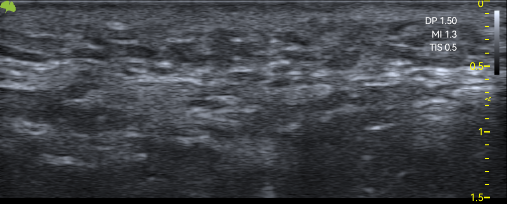 Real ultrasound image of the malar area: the same probe, eight different areas of the face, each with its own reading of the structure.
They are not competing technologies, they complement each other
A surface analyzer is still useful for what it does well: showing a patient their texture or spots in a visual, fast way with no preparation. But faced with a real clinical question (“which filler does this patient have, and since when?”, “is it safe to treat this area now?”) no camera analyzer can answer, because structurally it doesn’t see beyond the epidermis. That is where facial ultrasound comes in: it doesn’t replace the surface analyzer, it answers the question the surface analyzer can’t even ask.
If your clinic needs to confirm what lies beneath the skin before treating, not just how it looks on the outside, on the Nesai Derm page we explain what each report measures and which technical requirements your ultrasound machine needs. And if you want to go deeper into how a specific filler is detected, read how to tell which dermal filler you have.