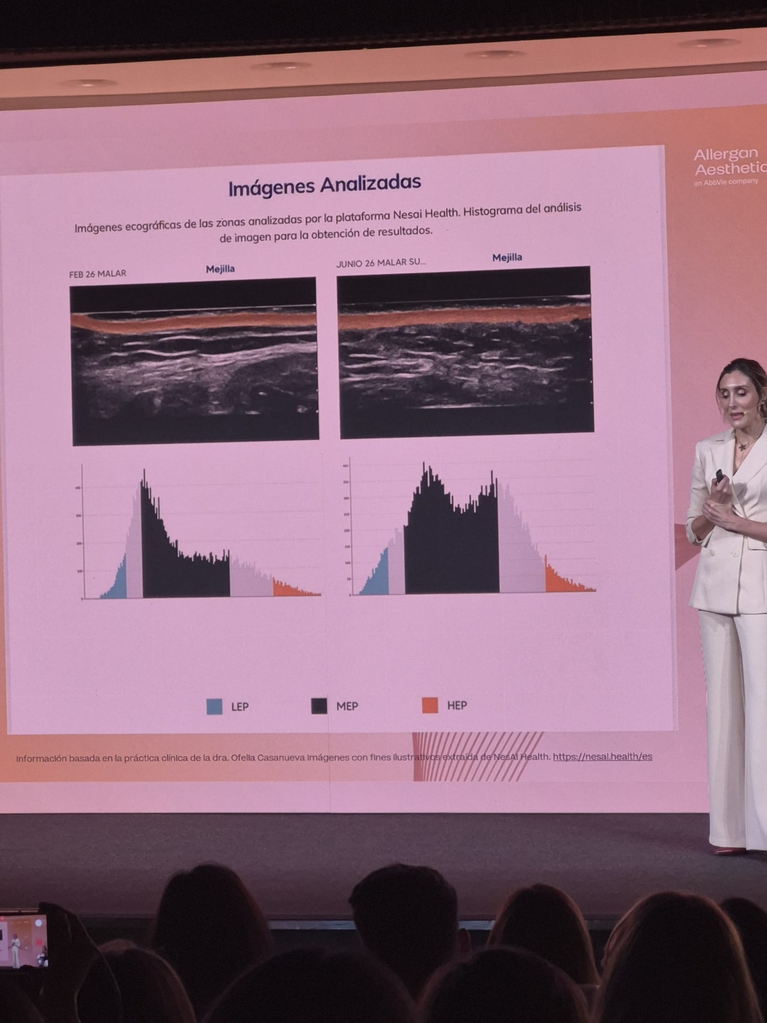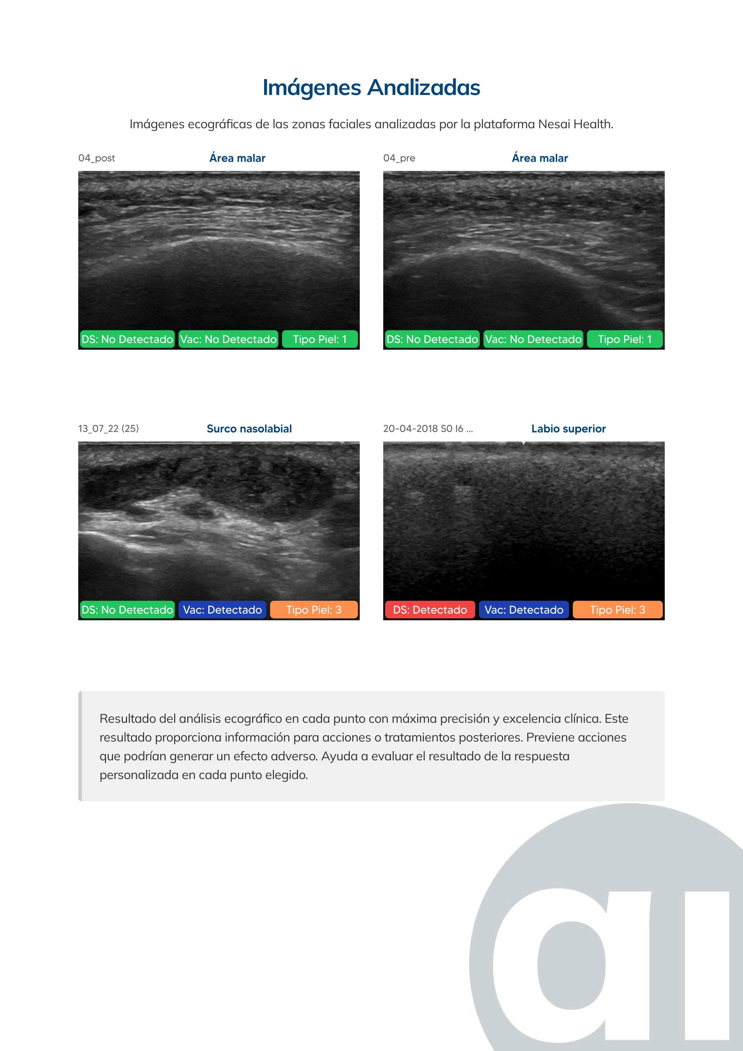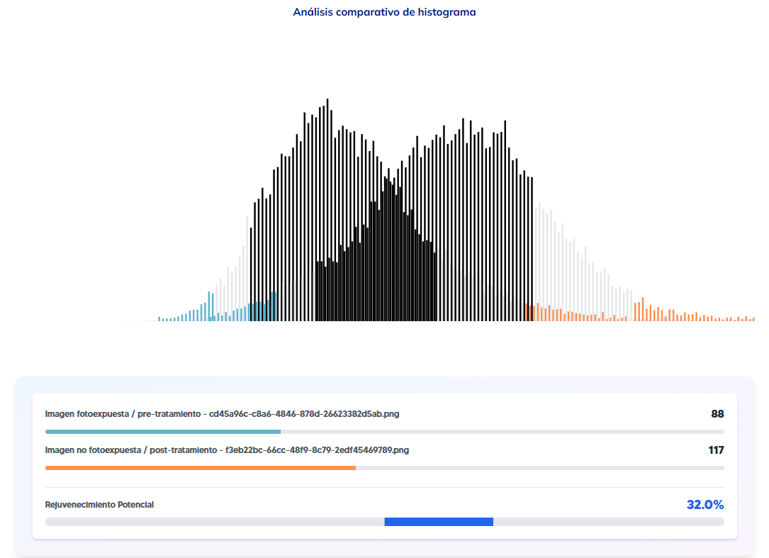
Can skin aging be measured with data, and not just by eye? Yes, and it isn’t a visual impression: it is structural data. Collagen, the extracellular matrix and elastosis change the density and echogenicity of the dermis in a way a high-resolution ultrasound can record. Analysed with AI, that image becomes a parameter you can compare between visits: not a subjective impression that “the skin looks better”, but a figure you can repeat in three, six or twelve months and compare with the previous one.
What happens in the skin as it ages
Skin aging combines two processes that are well documented in dermatology: intrinsic (chronological) aging and photoaging (from accumulated sun exposure). Both gradually break down the collagen and elastin in the dermis, reduce its density and disrupt its organisation: clinically, this shows up as loss of firmness, deeper wrinkles and skin that holds less water. The process isn’t fully visible to the naked eye until it is advanced; in the ultrasound image, however, damaged dermis has a different echogenicity from healthy dermis, even in early stages.
The parameters a facial ultrasound with AI measures
On the same ultrasound image, an AI system can identify and quantify:
- Collagen: density and organisation of the fibres in the dermis.
- Hydration: water content in the skin layers.
- Dermal thickness and density: the thickness of the dermis, which decreases with age and photoaging.
- SLEB (subepidermal low-echogenic band): a marker associated with accumulated sun damage.
- Elastosis: specific breakdown of collagen and elastin from UV exposure.
- Inflammatory processes: which can speed up or mask other structural changes.
None of these parameters can be detected with a camera-based skin analyzer, because they sit beneath the surface of the skin.
Why this is useful in practice, not just in theory
For an aesthetic medicine or dermatology clinic, having these parameters as objective data changes two things. First, it lets you justify with data why a specific treatment is recommended (collagen biostimulation, deep hydration, protection against photoaging) instead of relying only on the practitioner’s visual assessment. Second, it lets you measure the real effect of that treatment across successive visits, comparing the same parameter before and after, on the same area, with the same method.
The data supports the physician, it doesn’t replace them
The Nesai Derm report doesn’t need medical validation because it provides objective data about the dermis. The practitioner interprets it and decides on the treatment: Nesai gives them reproducible data that visual examination, by definition, can’t offer.
See the full detail of these parameters and the technical requirements on the Nesai Derm page.