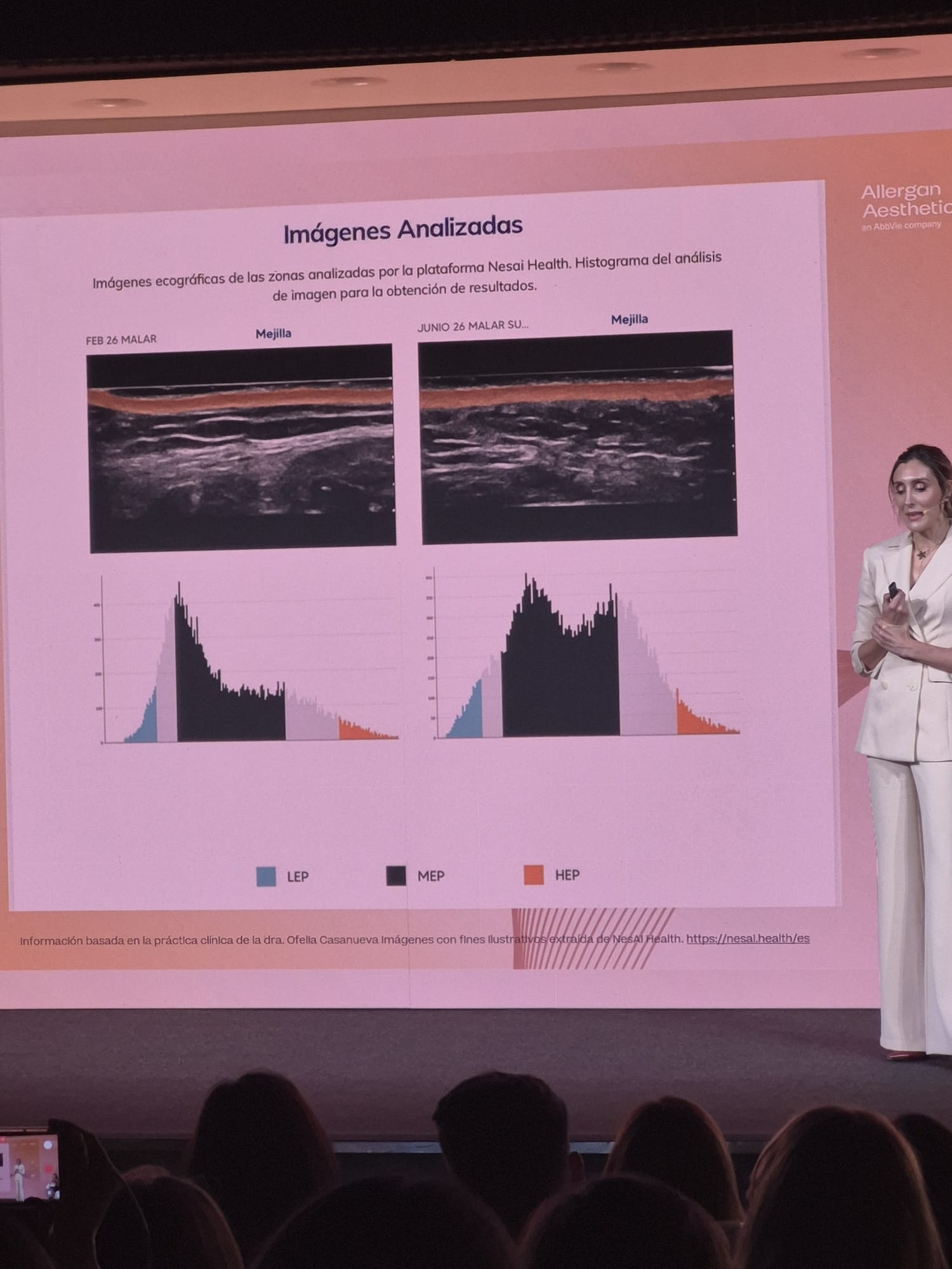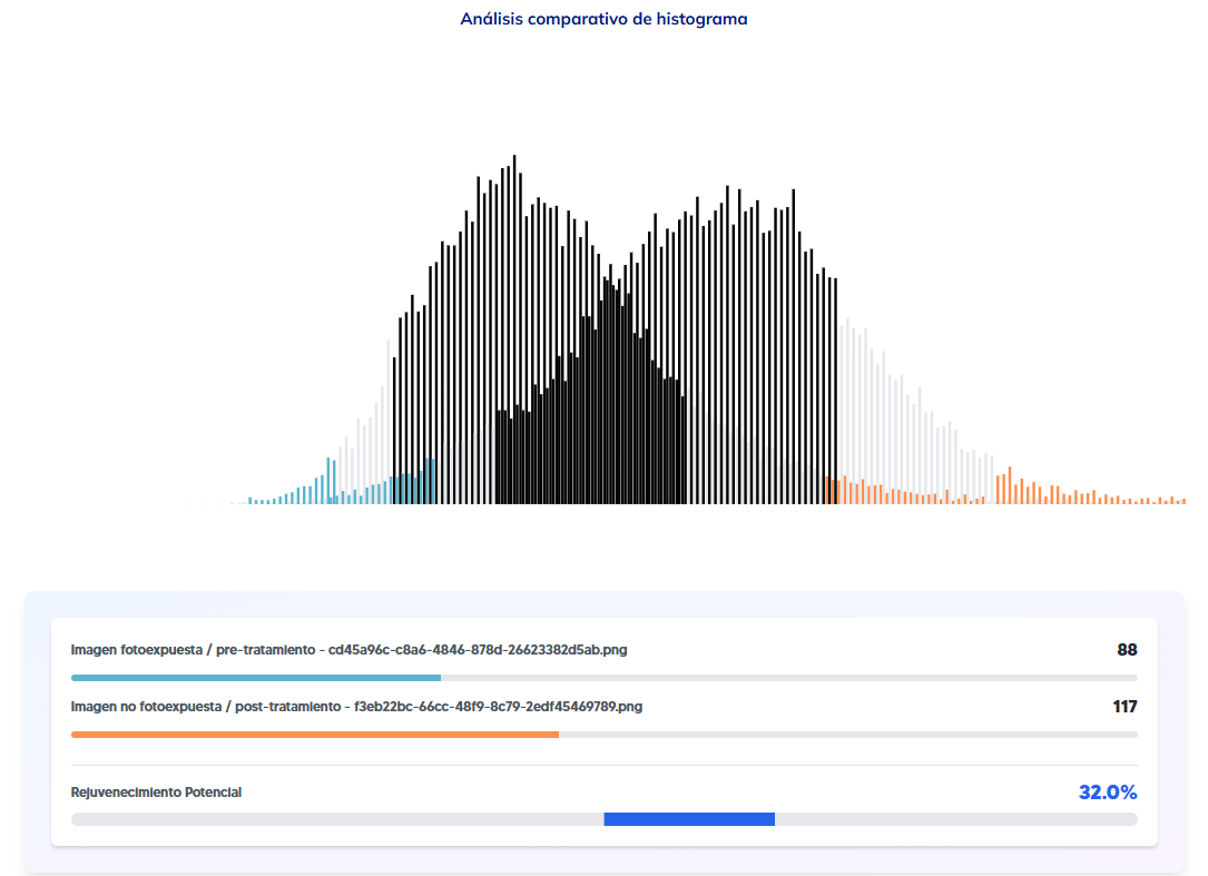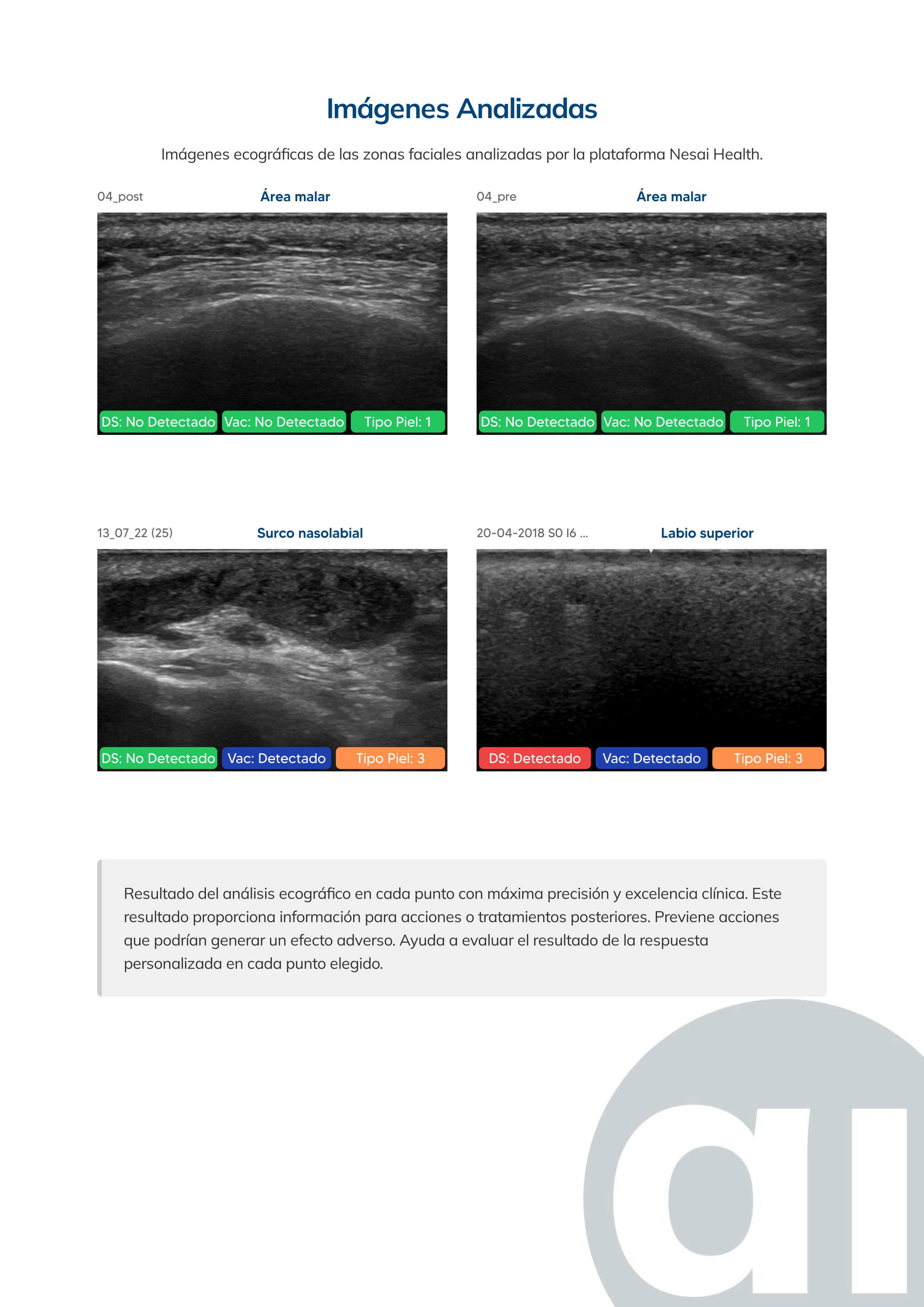
How can you tell which dermal filler someone has if there is no clear history? The answer isn’t in visual examination or touch: both can mistake a partially reabsorbed filler for inflammation, or miss a deep material altogether. The objective way to find out is high-resolution ultrasound: the ultrasound bounces back differently depending on the density and composition of the material it meets, which makes it possible to tell the patient’s own tissue apart from an injected filler, even years after the original treatment.
It is one of the most frequent questions in practice, and it keeps coming up more often: patients who change clinics, who don’t remember what was injected years ago, or who arrive with a doubtful assessment from a previous practitioner.
Why visual examination is not enough
A well-integrated filler can be impossible to tell apart from the surrounding tissue by sight or touch, especially after several years. The reverse is also a risk: treating an area on the assumption that a previous filler has fully reabsorbed, when residual material is still there, can lead to an overfilled look or to an adverse reaction when products are combined. Without objective data, the decision to treat rests on an estimate, not on a verified fact.
Which materials ultrasound can identify
The most common filler materials in aesthetic medicine behave differently in the ultrasound image, which makes it possible to tell them apart:
- Hyaluronic acid: the most common, with a characteristic ultrasound pattern.
- Silicone: non-absorbable, with a very well-defined ultrasound signal against the surrounding tissue.
- Calcium hydroxylapatite: a mineral, with a particular echogenicity due to its density.
- Biopolymers: not always authorised for clinical use, with an ultrasound pattern that helps tell them apart from conventional fillers.
Identifying which of these materials is present (and how much of it) is the basis for deciding whether it is safe to go ahead, wait, or refer to another type of treatment.
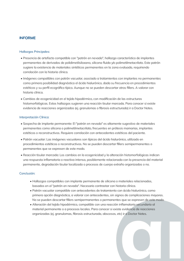 Findings page from a real Nesai Face report, identifying filler material present in the ultrasound image.
From the image to the clinical report
The practitioner performs the scan with their own clinic’s ultrasound machine. The image is analysed by AI in real time, and the system produces a preliminary report that a physician validates before it is shared with the patient. The data doesn’t replace clinical judgment: it supports it with something visual examination alone can’t offer.
Why this matters before a new treatment
Injecting without knowing what lies underneath is a clinical risk, for example when different products are combined. Confirming first which filler is there, how much is left and exactly where it sits makes it possible to adjust the treatment to the area that needs it, without overtreating an area that still holds more material than expected, or undertreating one that has already reabsorbed.
See what each Nesai Face and Nesai Derm report measures and which technical requirements your ultrasound machine needs.